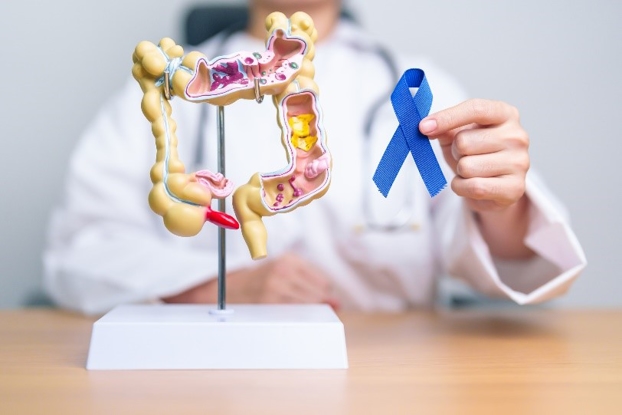
At Norman Regional Health System, we're committed to providing our patients
with exceptional and comprehensive gastroenterology care. Our experienced
gastroenterologists in Norman utilize state-of-the-art techniques and
therapies to diagnose and treat a wide range of digestive conditions.
Whether it's managing chronic diseases or providing diagnostic services,
trust that you will be in capable hands with our dedicated professionals.
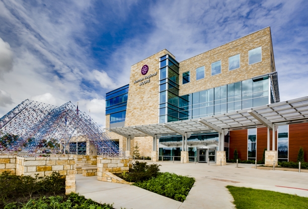
Comprehensive Gastroenterology Care & Advanced Imaging Services in
Norman and Moore
We focus on delivering the most accurate diagnoses and effective treatments
to manage and maintain your digestive health.
With personalized care and
advanced imaging services in Norman, we are able to diagnose and treat the following digestive disorders and more:
Celiac Disease: For those with celiac disease, ingesting gluten can cause significant
damage to the stomach lining. Symptoms often include diarrhea and fatigue.
Crohn’s Disease: This form of chronic inflammatory bowel disease can cause debilitating
symptoms, such as abdominal pain and severe diarrhea.
Diverticular Disease: Our team manages diverticulosis and accompanying symptoms with expertise.
Treatments may vary depending on symptoms and their severity.
Gallstones: We address painful gallstones that can cause intense upper abdominal pain.
GERD: We offer various treatments for the management of gastroesophageal reflux
disease (GERD) to alleviate discomfort from frequent acid reflux.
IBS: Irritable Bowel Syndrome (IBS) can severely disrupt daily life. Our approach
aims to minimize symptoms of stomach bloating and cramping to make daily
life more comfortable and manageable.
Peptic Ulcer Disease: Stomach pain from peptic ulcers requires careful management. Discuss a
personal management plan with your healthcare provider to determine the
best course of action for your daily needs.
Ulcerative Colitis: For ulcerative colitis (UC) patients, we offer treatments to manage long-lasting
inflammation and heal ulcers in the digestive tract.
Consult with a Gastroenterologist in Norman Today
For specialized gastroenterology care, we are here to provide precise diagnoses
and evidence-based treatment methods based on the individual needs of
each patient.
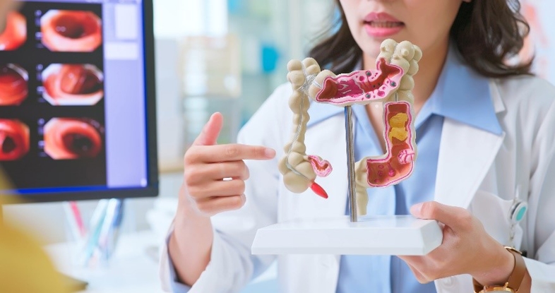
Diagnostic imaging performed by our Moore gastroenterologists may include:
Colonoscopy: A colonoscopy is a procedure performed to examine the inside of the rectum
and colon for any abnormalities.
Liver Biopsy: A liver biopsy is a procedure in which a small needle is inserted into
the liver to collect a tissue sample.
Capsule Endoscopy: Capsule endoscopies are procedures that use a tiny wireless camera to
take pictures of the digestive tract.
Esophagogastroduodenoscopy (EGD): An EGD is a procedure where a long, flexible tube, called an endoscope,
is used to examine the interior lining of the esophagus, stomach, and
duodenum (first portion of the small intestine).
Endoscopic Retrograde Cholangiopancreatography (ERCP): An ERCP is a highly sophisticated technique requiring special endoscopic
training where a long, thin, and flexible tube is used to enter the duodenum
and view the bile ducts.
Consult with a gastroenterologist in Norman today regarding your symptoms
and which diagnostic tests may be best recommended for your needs. Plan
your visit online or give us a call at
405-515-2777.
 GastroenterologyView Profile


.png)